Yeast contributions to Alzheimer’s Disease

Jamieson B Mcdonald, Sudip Dhakal, Ian G Macreadie*

School of Science, RMIT University, Bundoora, Victoria, Australia


Alzheimer’s Disease is a highly prevalent, age-related, neurodegenerative disease associated with the accumulation of toxic proteins, including amyloid beta and tau, that affect important cellular functions. Through the study of these proteins in yeast over the past 2 decades, the effects of amyloid beta oligomerization and aggregation, and tau hyperphosphorylation on basic cellular functions, such as ageing, oxidative stress, cell cycling and proteostasis have been observed. Strategies for the prevention of damage by these proteins can be explored, thanks to the exquisite array of technologies available for yeast studies. This review summarises existing knowledge of Alzheimer’s Disease, the work over the past two decades on yeast models for Alzheimer’s Disease and how these models contribute to the development of treatments and preventative strategies for Alzheimer’s Disease.


Globally, 50 million cases of cognitive impairment are associated with Alzheimer’s disease (AD), the most common form (60-80%) of dementia and major cause of death in elderly people. This number is expected to triple to 152 million by 20501,2,3. In addition to AD’s profound impact on quality of life, the disease is also a major healthcare, economic and social burden. It was reported in America alone, in 2018, an estimated 18.5 billion hours of care was provided by unpaid care workers and family members for those suffering from AD2. This care increases the risk of emotional distress and adverse mental and physical health consequences for family members and healthcare workers, which is valued at approximately $234 billion in the US alone2. Long-term care, health care, and hospital services for people over the age of 65 with dementia accounted for an estimated $290 billion in 20192. The associated healthcare costs of the disease are estimated to exceed $1 trillion by 20504.

AD mostly affects people above the age of 65 (late-onset AD), however, people as young as 40-50 years can develop genetically inherited, early-onset familial AD1. AD is a progressive neurodegenerative brain disorder that starts to develop years before symptoms arise2. During AD progression, disrupted cellular functions, loss of cellular homeostasis, and impaired cellular defenses cause significant irreversible structural and functional impairment to healthy brains leading to the death of neurons1,3. Damage to the hippocampus and neocortex leads to memory loss, language difficulties and learning defects4. Further, damage as AD progresses may result in a decline in other cognitive areas resulting in the complete incapacity to perform essential daily activities and the ability to function independently4.

AD is characterized by the extracellular accumulation of toxic amyloid beta (Aβ) plaques and intracellular hyperphosphorylation of tau in neuronal cells, oxidative stress, mitochondrial dysfunction, and neuronal atrophy1,3. Research has led to the development of several hypotheses to address the molecular mechanisms of AD5. However, the connections between these factors and neurodegenerative disorders remains elusive and the roles of the complex components of the disease has been disputed for decades. Despite, improved understanding of AD pathogenesis through the utilisation of yeast models and mammalian studies, gaps remain regarding the exact molecular changes and biological processes in the brain that lead to AD. These knowledge gaps present significant challenges in developing effective AD treatments. Presently there are no therapeutics prescribed for the prevention or alleviation of AD progression despite ~200 clinical trials in the past two decades searching for treatments.

The amyloid beta hypothesis represents the leading explanation for AD pathogenesis thus there is widespread research to inhibit Aβ-induced damage and subsequent death of neuronal cells6. Aspects of AD can be studied using numerous methods including ex vivo and in vitro experimental tools, computational studies, and transgenic models5. Approaches to reduce Aβ-induced AD focus on the inactivation or removal of Aβ via antibodies, and strategies to block the formation of toxic oligomers and plaques7. Translational outcomes have been constrained, owing to the shortcomings of animal models and their assay methods, including the high throughput screening of potential drugs and chemo preventatives7. There is, therefore, a need to adopt models that enable high throughput screening methods to accelerate the discovery and development of new treatment strategies. Yeast as a eukaryotic model organism has been utilized to address such limitations and has clearly demonstrated its effectiveness in studying the molecular aspects of AD.

This review article focuses on yeast’s pioneering contributions to AD research, and the recent insights and developments yeast has played in the study of AD. Emphasis is placed on the significant contributions yeast has made to translational research in the pre-screening of compounds that could be used to treat and/or prevent AD. Additionally, the review will highlight the way forward in using yeast model systems to provide further insights on the mechanisms of AD and for identifying multifactorial drugs or therapeutics that target AD.

The budding yeast, Saccharomyces cerevisiae, is the most popular unicellular model organism and has been used as a model for most AD studies described here. It was the first eukaryotic organism to have its genome fully sequenced and the information derived has facilitated substantial knowledge on cell biology relevant to humans. S. cerevisiae contains 6000 genes and ~31% have a homolog in humans4,8. The short generation time of yeast, ease of culture and its ability to be readily transformed to produce human proteins in a desired context make it an efficacious model for studying chronic diseases such as AD. Although yeast lack specific processes of neuronal cells, a nervous system, immune system, and neuropathology associated with cell-cell communications, the majority of the molecular signaling pathways and proteins involved in human neurological diseases exhibit conserved sequences and function9,10. A large fraction of essential cellular processes, including cell cycle progression, mitochondrial biology, metabolism, transcription, translation, protein secretion, protein quality control mechanisms, vesicle trafficking and apoptosis are highly conserved between this unicellular fungus and humans, including our neuronal cells4, providing an efficacious platform to study protein-misfolding pathologies11. Hence, S. cerevisiae provides a facile means of studying both physical and genetic associations between proteins and simple cellular mechanisms, paving the way for the utilisation of yeast in medicinal and medical research. Yeast have played an essential role in the development of many high-throughput techniques including DNA and ChIP-chip microarrays, in the large scale analysis of protein-protein interactions (yeast two-hybrid and three-hybrid systems), transcriptomics, proteomics and metabolomics analyses9.

Ageing is an inevitable factor of life and an underlying cause of numerous chronic diseases such as AD. Ageing is a multifactorial process involving the build-up of molecular, cellular, and systemic damage leading to physiological impairment and a decline in reproductive ability and survival12. Ageing humans exhibit very similar features to ageing S. cerevisiae (that are identified by 2 or more bud scars) such as accumulation of aberrant proteins, reduced telomeres, oxidative damage and mitochondrial dysfunction10. An advantage of using yeast for AD research is that it is less complex compared to mammalian models and affords a relatively simple means to investigate elements associated with AD. In addition, the use of S. cerevisiae enables large scale genetic screening and functional genomic studies that are not achievable in humans. Furthermore, because the budding yeast displays similar cellular aspects of ageing humans it can be utilised as a model to screen compounds that reinstate health to old cells and to identify potential therapeutics. As a result, yeast models have provided unprecedented insights into the underlying molecular basis of ageing and to decipher the complexity of disease pathology and disease associated mutations such as those involved in AD.

All the features discussed above make yeast an efficacious system for genome-wide screening of genes that affect lifespan or the toxicity of heterologously expressed human proteins such as Aβ and tau. In addition, the investigation of gene and protein interaction networks regulating longevity and ageing are achievable. Yeast have been extensively utilized as a model organism for studying AD (Figure 1) and neurodegeneration as discussed in some previous reviews4,5,7,10.

JHCG-20-1114-Fig1

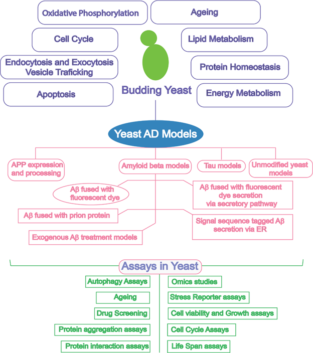
Figure 1: Schematic diagram outlining the biological similarity of yeast and research tools offered by yeast to enable exploration of yeast as a model for Alzheimer’s disease. AD, Alzheimer’s disease; APP, amyloid precursor protein; Aβ, amyloid beta; ER, endoplasmic reticulum.

Although Aβ and tau have no orthologs in yeast, their heterologous expression is readily achieved and can be highly informative in understanding the pathobiology of these proteins4. Yeast models have contributed to understanding the pathology of the disease via modelling amyloid precursor protein (APP) processing, in vivo Aβ oligomerization, Aβ-associated toxicity, and tau phosphorylation10. Table 1 shows the contributions of yeast cell-based assays to the study of AD and recent advances in the use of yeast.

Table 1. Yeast AD Models with main findings and insights

Amyloid Precursor Protein (APP) expression and processing models

  • Human γ-secretase activity reconstituted in yeast13 generated Aβ40, Aβ42 and Aβ4314.
  • α-secretase activity modeled in yeast15.
  • Yeast proteases Yap3 and Mkc7 exhibit α-secretase activity which act on APP in the late Golgi16.
  • Yeast two-hybrid system screened proteins that interact with APP C-terminal domain including phosphotyrosine-binding domain proteins, Gulp1 and staufen117,18,19.
  • Amyloid beta models
  • A yeast two-hybrid system revealed Aβ interacts with itself in vivo20.
  • 42 (fused to GFP) caused cell stress and a heat shock response, slowing growth of cells21.
  • Aβ oligomers and fibrils caused stress responses to yeast mCherry fluorescence reporter22.
  • 42 fused to GFP aggregated, was toxic and induced autophagic responses, allowing screening for chemo preventatives7.
  • 42-GFP fluorescence levels were reduced by folinic acid23 and simvastatin24. Simvastatin also reduced native Aβ42 levels24. 
  • Latrepirdine (Dimebon™) enhanced autophagy and reduced intracellular GFP-Aβ42 levels25.
  • Nutrient and stress signaling genes (RRD1, SNF4, GCN4 and SSE1) involved in the clearance of GFP-Aβ aggregates26.
  • 42 increased cytotoxicity, ROS, proteasomal activity and mitochondrial dysfunction27.
  • 42 toxicity showed a synergistic interaction between energetics and ER stress28. Aβ42 expression resulted in reduced cell growth rates and yields demonstrating a switch of energy from growth to maintenance, triggering ER stress28.
  • 42 caused feedback inhibition of presequencing processing and to the accumulation of mitochondrial preproteins and preprocessing enzymes. Mitochondrial Aβ42 inhibited precursor maturation, causing mitochondrial dysfunction, increased levels of ROS and decreased respiration and membrane potential29
  • Aβ toxicity can be examined with chemicals like tyramine to investigate toxic synergy between certain compounds and Aβ. Tyramine enhanced Aβ toxicity causing elevated ROS production, mitochondrial damage, and respiratory growth inhibition30.
  • 42 oligomerizes and aggregates in vivo. Replacement of the [PSI+] prion forming domain with Aβ42 restores the protein’s ability to aggregate31.
  • Fusion of Aβ42 with MRF (Sup25p lacking N-terminal domain) caused aggregation into oligomers, deeming Sup35 unable to terminate translation32.
  • FMN1, responsible for riboflavin kinase expression, and its metabolic product flavin mononucleotide (FMN) reduced Aβ toxicity33.
  • Two inhibitors of Aβ42 oligomerization found in a library of 12,800 drug-like small molecules34. Seven drugs from 1,200 FDA approved drugs which reduce Aβ42 oligomerization identified. Overexpression of Aβ toxicity modulator Yap1802 reduced Aβ42 oligomerization35.
  • Chemically synthesized exogenous Aβ42 was toxic to yeast36. Fibrils were not toxic36. Role of fibrils and oligomers in the propagation and toxicity of Aβ42 demonstrated.
  • Proline-rich whey peptides inhibited Aβ42 oligomerization via suppression of β-sheet and anti-parallel β-sheet structures, also demonstrate to dissociate pre-existing β-sheet structures in Aβ42. Suppression of structures was associated with rescue of yeast cells and SH-SY5Y neuronal cells37.
  • Rapid absorbance-based growth assay to screen the toxicity of Aβ42 and protective agents against cell death38.
  • Exogenous 42 caused growth of yeast in stationary phase, suggesting unscheduled cell cycling is stimulated by Aβ4239.
  • 42 rescued Candida glabrata from hydroxide toxicity, which increased cell survival40.
  • 42 fused to the C-terminus of the ER targeting signal Kar2 resulted in toxicity towards yeast cells11.
  • Constitutively expressed native Aβ42 resulted in a lower cell growth rate, biomass yield, respiratory rate, proteasomal activity and increased oxidative stress27.
  • Galactose-induced expression of eGFP-Aβ42 possessing a TEV cleavage site demonstrated mitochondrial Aβ42 induces mitochondrial dysfunction via inhibition of preprotein maturation29.
  • Overexpression of Yap1802 moderately restored endocytosis in yeast cells and suppressed Aβ42 toxicity: validated in in C. elegans and rat hippocampal neurons11.
  • Clioquinol reduced levels of Aβ via the degradation of Aβ oligomers, restoration of endocytosis and amelioration of Aβ toxicity41.
  • Clioquinol and dihydropyrimidine-tiones act via a metal-dependent mechanism which alleviated Aβ toxicity via increased Aβ turnover, restoration of vesicle trafficking and protection against oxidative stress42.
  • 42 fused to GFP and an ER targeting signal demonstrated intracellular traffic pathways are essential to produce toxic species and yeast PICALM ortholog (Yap1802) participates in cellular toxicity. Aβ42 indicated to cross intracellular membranes and impact mitochondrial organelles43.
  • Tau biology models
  • Tau isoforms 3R and 4R indicated processes of tau phosphorylation and aggregation are regulated by yeast kinases Mds1 and Pho85 (orthologs of human tau- kinases GFK-3β and cdk5, respectively44.
  • Hyperphosphorylation of tau and binding to microtubules was found to be inversely proportional to one another: microtubule instability is caused by aggregation of tau on microtubules45.
  • Phosphorylation at tau site S409 was found to moderate tau aggregation and microtubules. Oxidative stress and mitochondrial dysfunction found to enhance tau aggregation46. Peptidyl prolyl cis/trans isomerase human Pin1 and its yeast ortholog Ess1 indicated to participate in tau phosphorylation and structural assembly. Accumulation of hyperphosphorylated and aggregation-prone tau found to cause cytotoxicity47.
  • α-synuclein and tau found to cause a synergistic toxicity to yeast cells48. Expression of α-synuclein and tau resulted in elevated tau phosphorylation and aggregation49.

APP, a single-pass transmembrane protein present at high levels, is metabolised in a rapid and multifaceted manner by γ-secretase and β-secretase, or α-secretase50. APP processing via α-secretase prevents the production of toxic Aβ peptides. However, the proteolytic cleavage of APP by β-secretase and γ-secretase results in the aggregation of toxic, highly amyloidogenic and hydrophobic peptides including Aβ40, Aβ42, and less abundant Aβ4350. The amino acid sequences of APP gene present at Chromosome 21 vary due to variations and mutations (Figure 2). The mutations in coding sequences sometimes are pathogenic and among these some are implicated in early-onset AD (Figure 3).

JHCG-20-1114-Fig2

Figure 2: Detailed region of chromosome 21 showing amyloid precursor protein (APP) location, protein families and variations in the APP gene51.

JHCG-20-1114-Fig3

Figure 3: Amino acid changes leading to the early onset of Alzheimer’s disease.

The amino acid sequence of Aβ is given at the top. Amino acid changes in FAD have been highlighted (red = pathogenic, green = protective): Swedish mutation, a double mutation, substitution of lysine and methionine with asparagine and leucine which amplifies Aβ production52. London mutation, substitution of valine to isoleucine at position 717 of APP53, increases Aβ42/Aβ ratio54. Indiana mutation, missense mutation causing replacement of valine with phenylalanine55. Flemish mutation, substitution of alanine with glycine at position 2156, causes aggregation and fibril generation in brain blood vessels57 Dutch mutation, the most common, replacement of glutamic acid with glutamine at position 22, causes severe amyloid deposition and premature death58; Iowa mutation, aspartic acid replaced with asparagine at position 2359, promotes Aβ fibrillogenesis and causes an increase in Aβ-induced toxicity60. Arctic mutation, missense mutation causing replacement of glutamic acid with glycine at position 22, causing enhanced protofibril formation61. Piedmont mutation, missense mutation causing replacement of leucine with valine at position 34, causes deposition of Aβ fibrils62. Italian mutation, replacement of glutamic acid with lysine at position 22, causes Aβ immunoreactivity63. Osaka mutation, deletion of glutamate at position 22, enhances Aβ oligomerization64. English mutation, histidine replaced by arginine at position6, accelerates Aβ fibril formation65. Taiwanese mutation, replacement of aspartic acid with histidine at position 7, increases production of Aβ40 and Aβ42, Aβ42 is preferentially increased66. Tottori mutation, replacement of aspartic acid with asparagine at position 765, alters assembly kinetics of Aβ67. Leuven mutation, replacement of glutamic acid with lysine at position 11, elevates Aβ42/40 ratio, increasing total Aβ levels68. Iranian mutation, replacement of threonine with alanine at position 4369. Austrian mutation, replacement of threonine with isoleucine at position 43, significantly increases Aβ42/40 ratio, elevating Aβ42 secretion and decreasing Aβ40 secretion70. German mutation, replacement of valine with alanine at position 715 of APP, increases Aβ42/40 ratio71. Iberian mutation, replacement of Isoleucine with phenylalanine of position 716 of APP, significantly elevates Aβ42/40 ratio72. Australian mutation, replacement of leucine with proline at position 723 of APP, increases levels of secreted Aβ73. Belgian mutation, replacement of lysine with asparagine at position 734 of APP, increases Aβ42/40 ratio74. Icelandic mutation, replacement of alanine with threonine at position 2175, protects against AD via a reduction amyloidogenic peptides76.

Yeast-based systems have provided valuable insights into APP processing, but in this review, APP is only discussed briefly with greater emphasis placed on yeasts contributions to the study of Aβ and tau. Yeast APP models have been utilised to study APP-like substrates and secretases related to the processing and formation of Aβ13,14,15,16. Models have demonstrated yeast proteases Yap3 and Mkc7 exhibit α-secretase activity which acts on APP in the late Golgi bodies16. Studies have reconstituted γ-secretase activity in yeast, which was revealed to generate Aβ40, Aβ42 and Aβ4314. The yeast two-hybrid system has been utilised to identify proteins which interact with the intracellular cytoplasmic domain of APP17,18,19. Using a yeast γ-secretase assay, specific mutations in presenilin 1 were shown to cause conformational changes in γ-secretase which regulated Aβ trimming, demonstrating a relationship between PS1 conformational changes and γ-secretase activities77. Further analysis of APP mechanisms in yeast will provide important information on the mechanisms of Aβ production and potential targets to inhibit Aβ generation.

It has been well established that an imbalance involving the assembly and clearance of amyloid beta and related Aβ peptides is an early, clear and often initiating factor of AD6. Despite various isoforms of Aβ, with differing capacities for aggregation, three main groups of Aβ assemblies exist78 which include monomers, higher soluble oligomers, and insoluble fibrillary polymers3,78. The most abundant peptide produced in the brain is Aβ40 followed by Aβ42, which are predominately localized extracellularly of brain cells3,78. The production of monomeric Aβ is followed by a sequence of reactions which form oligomers and fibrils. Elevated levels of Aβ42 with an increased ratio of Aβ42:Aβ40 leads to the formation of plaques and deposits, which act as reservoirs for toxic Aβ peptides3. The formation of these structures results in microglial activation, oxidative stress, mitochondrial dysfunction, inflammatory responses, synapse dysfunction, and interferes with cellular communications, resulting in a cascade pathway to disease and neuronal atrophy3,4,6.

Oxidative stress is inextricably linked to the amyloid beta hypothesis. The brain exhibits the greatest rate of oxidative metabolism. Neuronal cells are more susceptible to reactive oxygen species (ROS) and free radicals owing to high oxygen consumption and deficiency in antioxidant enzymes available in the brain compared to other organs2. The generation of ROS may be one of the initial factors contributing to the development of AD following the increased deposition of Aβ42. ROS are produced via several pathways; a significant player contributing to the production of ROS are mitochondria2,30,79. ROS produced by mitochondria can result in the impairment of mitochondrial membrane potential, leading to further ROS production resulting in mitochondrial dysfunction, eventually causing oxidative damage to whole neuronal cells79. The hydroxyl radical, being the most reactive one, contribute to oxidative damage on both the Aβ peptide itself and on surrounding molecules (proteins, lipids and DNA)30. Therefore, evidence indicates that Aβ42 and its interplay with modified mitochondrial function and energy metabolism exacerbates the progression of AD and represents an early event in AD-affected brains27. Apart from the increased production of ROS, impaired mitochondrial function can constitute disrupted electron transfer, ATP synthesis, mitochondrial transcription, mitochondrial pre-protein maturation and protein synthesis27. However, the mechanisms of mitochondrial dysfunction and Aβ play in AD pathogenesis remains elusive. Yeast models have made several contributions to understanding the pathology of the disease via several approaches that focus on in vivo Aβ oligomerization, Aβ-associated toxicity, oxidative stress and mitochondrial dysfunction10.

Yeast has been utilised to address unknown questions and to improve understanding regarding in vivo Aβ oligomerization, aggregation, toxicity, and the intracellular pathological dynamics of the peptide4,5,7,10. One of the earliest developments used a yeast two-hybrid system (Aβ linked to the LexA DNA-binding domain (bait) and as well as to transactivation domain (prey)20. This initial study was motivated by the idea that Aβ was capable of interaction with itself in vivo. The expression of these two fusion proteins in S. cerevisiae allowed protein-protein interactions to be evaluated which demonstrated the specific nature of Aβ self-interaction as replacement of Aβ with a Drosophila protein resulted in no interaction20. This effective use of the two-hybrid system also identified amino acids of Aβ involved in its self-interaction and subsequent nucleation dependent aggregation.

The majority of Aβ produced is extracellular, therefore its impacts on cellular viability can be ascertained by adding chemically-synthesized Aβ to cells5. To demonstrate extracellular Aβ toxicity towards yeast cells a study showed cell death when exposed to chemically synthesized Aβ in water36. The yeast Candida glabrata was used in these studies because of its ability to form colonies overnight on solid media. Aβ was toxic when freshly prepared, but fibrillar Aβ was not toxic36. This yeast model has been implemented to explore the neuroprotective properties of whey derived peptides. This approach demonstrated proline-rich whey peptides (wPRP) could inhibit Aβ oligomerization via suppression of β-sheet and anti-parallel β-sheet structures, while also having the ability to dissociate pre-existing β-sheet structures in Aβ37. Suppression of these structures was shown to be associated with the rescue of yeast cells and SH-SY5Y neuronal cells indicating an important association between β-sheet structure and oligomer toxicity37.

The assays discussed above are based on colony counting methods and are laborious and inappropriate for screening gene deletion and overexpression libraries as well as small molecule collections. Bharadwaj and colleagues adapted the method discussed above by developing a rapid absorbance-based growth assay to screen the toxicity of Aβ and protect against cell death38. In contrast to the studies above, S. cerevisiae was the model organism used instead of C. glabrata. The colony forming unit assay previously developed and the absorbance-based growth assays were compared both revealing oligomer Aβ toxicity adopts a dose dependent toxicity profile38. The assay developed was tested with autophagy enhancers (latrepirdine, rapamycin and SMER28), demonstrating protection of yeast and mammalian cells from oligomer Aβ-induced cell death: each of the autophagy enhancers were shown to be crucial for protection against Aβ toxicity38. The yeast absorbance viability assay developed represents a simpler technique than colony forming assays with greater sensitivity for screening gene deletion libraries, expression libraries and compound repositories. Additionally, it further validates the protective ability of autophagy inducers (providing evidence for autophagy manipulation as having positive therapeutic implications against Aβ mediated cell death. This large-scale screening of compounds that can rescue or protect cells from Aβ-induced cell death provides an efficient approach to the discovery of potential chemo preventatives for AD treatment.

The complexity of studying toxicity of extracellular Aβ is well documented. Varying methods of Aβ preparation have been shown to result in differences in its biological activity. For example, Porzoor and colleagues demonstrated that yeast in stationary phase growth treated with extracellular chemically-synthesized Aβ prepared in ammonium hydroxide caused growth proliferation39. The study addressed an important observation that brains affected by AD exhibit terminally-differentiated neurons that have entered a cell cycle, but then die as a result. It has been suggested that unscheduled cell cycle entry is stimulated by Aβ5. Thus, results from the latter study provide insights into yeast possibly being triggered in the same way. However, in contrast to neuronal cells, yeast in stationary phase have substantial intracellular reserves enabling them to complete one or two cell divisions in the absence of added nutrients5.

Exogenous Aβ treatment has also been shown to rescue yeast from hydroxide toxicity40. Sodium hydroxide (NaOH) has been shown to solubilize exogenous synthetic Aβ however, Dubey and colleagues revealed exogenous Aβ had a strong protective effect on C. glabrata cells against NaOH toxicity, increasing cell survival40. The protective mechanism observed may be that the interaction between Aβ and C. glabrata cell membrane protects periplasmic and membrane proteins from NaOH-induced denaturation40. Results from this study provide evidence that both the physiological and pathological roles of Aβ depends on numerous factors.

Aβ can be found inside cells, in locations including the cytosol, secretory pathway, autophagosomes, and mitochondria5. The heterologous expression of Aβ in yeast cells, allows the development of new approaches and yeast-based models to monitor the presence of intracellular Aβ and its toxicity. These systems have been coupled with genome wide association studies (GWAS) to identify AD risk factors. The systems developed offer a significant advantage in studying Aβ pathology as they limit the amount of heterologously expressed protein and, thus, also any unwanted non-Aβ-associated pathologic side reactions4. Additionally, these systems coupled with an array of technologies provide a high throughput means of identifying genes and compounds which may reduce or exacerbate Aβ toxicity. Innovative and recent developments in yeast model systems involving the heterologous expression of Aβ are discussed below.

The generation of Aβ takes place in the plasma membrane and processing of the toxic peptide can therefore involve the secretory pathway, resulting in the release of Aβ into trans-Golgi, endosomal compartments, and extracellular space5. This can result in the secretion and re-uptake of the peptide in cells via multi-compartment trafficking and as a result Aβ can interact with the plasma membrane, undergo endocytosis and vesicular trafficking11. As a principal organelle responsible for protein quality control and secretion, the endoplasmic reticulum (ER) is significantly disrupted in AD neurons33. Elevated production of Aβ and subsequent aggregation has been suggested to result in abnormal levels of ER stress, playing a role in synaptic dysfunction in AD33.

A pioneering study performed by Treusch and colleagues formulated a yeast model system which involved the production of Aβ fused to the C-terminus of the ER targeting signal Kar2 (known as ssAβ 1-42). This approach enabled Aβ to be targeted to the ER and released into the secretory pathway, where it could be subjected to endocytosis, and thus transported via endocytic compartments potentially associated with AD11. Expression of ssAβ 1-42 resulted in a decrease in yeast cell growth. Additionally, strains with uniform stable ssAβ 1-42 expression were obtained by integrating tandem copies of the fusion protein into the genome thus, permitting yeast screening to identify genetic suppression. A genome-wide screening of >5000 ORFs revealed several suppressors and enhancers of Aβ toxicity4,11. Modifiers were involved in receptor-mediated endocytosis11. For example, overexpression of Yap1802 gene, the yeast ortholog of the human AD risk factor, phosphatidylinositol binding clathrin assembly protein (PICALM) moderately restored endocytosis in yeast cells and suppressed Aβ toxicity11. These observations were validated in C. elegans and rat hippocampal neurons in which PICALM expression partially prevented Aβ-induced cell death11.

This yeast model assay system has been further utilised in high throughput screens to identify compounds that prevent or rescue Aβ-associated cell toxicity. For example, clioquinol was demonstrated to reduce Aβ via the degradation of Aβ oligomers, restoration of endocytosis and amelioration of Aβ toxicity5,41. Tardiff and colleagues validated clioquinol’s ability to alleviate Aβ toxicity using a phenotypic small molecule screen: in addition, dihydropyrimidine-thiones (DHPM-thiones) also reduced Aβ toxicity. Both clioquinol and DHPM-thiones acted via a metal-dependent mechanism which rescued cells from Aβ toxicity. Cotreatment of yeast cells with these compounds ameliorated Aβ toxicity via increased Aβ turnover, reducing Aβ levels and restoring functional vesicle trafficking42. Additionally, the synergistic interaction between these compounds protected cells from oxidative stress. Results of yeast studies were validated in a nematode expressing Aβ where single and combinatorial treatments of the two compounds reduced death of neurons42. This observation importantly indicates that expression of Aβ results in homogenous cellular pathologies from yeast to neurons. D’Angelo and colleagues slightly altered the yeast models utilised in the studies discussed above by fusing Aβ42 to GFP43, further demonstrating that intracellular traffic pathways are essential to produce toxic species and that yeast PICALM orthologs participate in cellular toxicity43. Yeast expressing Aβ-GFP disturbed the cell-traffic pathway resulting in a partial reduction in Aβ-GFP toxicity. In addition, the ability of Aβ42 to cross intracellular membranes and impact mitochondrial organelles was observed43. Further investigation of Aβ ability to impact organelles and its roles in induced mitochondrial dysfunction and cytotoxicity will be discussed further later.

It is important to note inconsistency in the results between the two studies41,43 may be due to varied expression levels of the different constructs. Further research is necessary to clearly establish the role of PICALM, endocytosis and their involvement in dealing with numerous levels of Aβ aggregation occurring during different stages of AD4,5. Both studies used inducible promoters for the expression of Aβ peptides, whereas Aβ production in neurons is constitutive33. Although, an inducible promoter enables favourable induction of acute cytotoxicity it is paralleled by significant change in carbon-source and metabolism33. These initial studies11,41,43 provided a great deal of knowledge regarding the ability of Aβ to re-enter cells through endocytosis, avoiding transit through the secretory pathway and exportation from the cytoplasm. Subsequently, Aβ peptides form more oligomers which results in an increased cellular toxicity in yeast. In addition, these studies provide important insights into genetic and compound-based targets that could be explored to reduce Aβ toxicity.

Following on from the above studies, Chen and colleagues33 used a different yeast model system and approach to study the effects of Aβ, observing reduced respiratory function, elevated levels of ROS and finding ways to alleviate toxicity. In contrast to the studies discussed above, yeast constitutively expressed Aβ42 and the model was coupled with a synthetic genetic array (SGA). This approach enabled Aβ-toxicity modifiers to be identified and investigated and the mechanistic interactions between genes and their related pathways permitted the mapping of genome-wide interaction networks implicated in Aβ toxicity. FMN1, the gene responsible for riboflavin kinase expression, and its metabolic product flavin mononucleotide (FMN) was discovered to reduce Aβ toxicity33. Gene set enrichment analysis demonstrated yeast mutants involved in protein secretion and degradation processes had increased sensitivity to Aβ toxicity. Defects in riboflavin metabolism resulted in increased Aβ toxicity and SGA screening indicated the yeast fmn1 mutant was sensitive to Aβ toxicity with complementation assays validating that the mutant was critical for Aβ toxicity33. The fmn1 mutant strain constitutively expressing Aβ when cultivated in media supplemented with FMN resulted in reduced Aβ toxicity. Results suggest that preserving adequate levels of FMN may be necessary for cell growth and that FMN deficiency could play a role in Aβ triggered toxicity.

Additionally, the study investigated whether the fmn1 mutant contributed to protein aggregation using GFP-tagged Hsp104 as a reporter for protein aggregation. Heat shock of the fmn1 mutant strain resulted in significantly greater misfolding and aggregation of the toxic peptide33. FMN supplementation was indicated to play an important role in protein misfolding as supplementation with FMN decreased Aβ aggregate formation. Challenging the Aβ-producing strain with endogenous ROS H2O2 allowed oxidative stress implications involved in AD to be assessed. Oxidative stress tolerance assays and growth assays determined H2O2 caused significant oxidative stress to the Aβ-producing strain (prolonged lag phase of growth). Supplementation with FMN improved cell viability and therefore, could be beneficial for oxidative stress tolerance, in both Aβ-producing and control strains: thus, FMN improves redox homeostasis33. The study examined the possible roles of flavoenzymes in Aβ toxicity by assessing strains with single flavoenzyme gene deletions. Coinciding with previous results, strains supplemented with FMN significantly increased cell viability in single gene mutant strains constitutively expressing Aβ. Overexpression of flavoprotein Dus2p improved viability in dus2 mutant expressing Aβ thus modulating and reducing Aβ toxicity. This discovery suggests DUS2 could be involved in regulating Aβ toxicity and should be investigated as an extra gene target together with FMN1 for reducing Aβ toxicity.

These studies further validate Aβ yeast models used in conjunction with GWAS and or SGAs as an effective and valuable approach to identify Aβ toxicity modifiers and their associated mechanisms. Continued application of this approach will prove to be highly beneficial in understanding the molecular mechanisms of AD, and for identifying and discovering appropriate therapeutic targets.

Further investigation of Aβ ability to impact organelles and its roles in induced mitochondrial dysfunction and cytotoxicity has been recapitulated in yeast. Chen and Petranovic utilising a slightly different approach27 to other studies35,43 which used inducible promoters resulting in a strong acute cytotoxicity, developed a yeast model that constitutively expresses human native Aβ possessing the ER signal sequence Kar2 for secretion, only partially effecting growth27. The production of Aβ monomers and oligomers in cells resulted in lower growth rate and biomass yields27. Additionally, mitochondrial dysfunction (lower respiratory rate) was observed to be directly associated with increased production of ROS (increased oxidative stress) and decreased proteasomal activities27. Decreased proteasomal activity was seen in all three proteolytic activities of the 20S proteasome suggesting that a surplus of misfolded proteins in Aβ strains disrupts the proteasome preventing it from degrading other substrates which may also contribute to cell cytotoxicity. Inhibition of proteasome systems has been suggested to play an important role in AD related dysfunction of the ubiquitin-proteasome system. Chen and Petranovic recently reviewed the role of frameshift ubiquitin B in AD80.

Recently a systems biology approach by Chen and colleagues demonstrated a synergistic interaction between energetics and ER stress increased Aβ toxicity28. The study provides further evidence on the complex nature of Aβ and how it is influenced by certain conditions. Utilising bioreactor cultures, for a more stable (controlled) environment, significant differences in the mechanisms associated with the cytotoxicity of Aβ40 and Aβ42 was revealed. The expression of Aβ40 induced mild stress whereas, expression of Aβ42 caused a sustained high ER stress response and an unfolded protein response (UPR) affecting mitochondrial function (lower respiration rates and reduced ATP generation), increasing levels of ROS resulting in reduced growth rates and biomass yields, a shorter chronological lifespan and an increase in lipid synthesis disrupting lipid metabolism28. The use of this yeast model system in a highly controlled environment provides an improved understanding of cellular responses to Aβ toxicity mechanisms and the associated stresses placed on cells. Chen and colleagues demonstrate that their yeast model system can be used to elucidate pathways involved in cytotoxicity but can also be modified for the screening of compounds that could potentially alleviate oxidative stress, UPS dysfunction and mitochondrial dysfunction.

Other studies have adopted a different approach to studying Aβ implications on mitochondrial dysfunction. Mossmann and colleagues demonstrated mitochondrial Aβ induces mitochondrial dysfunction via inhibition of preprotein maturation29. The study isolated mitochondria from a yeast mutant that lacks Cym1 (PreP homolog) to analyse the physiological impact of peptide turnover on mitochondrial functions29. Mitochondrial presequence processing depended on peptide turnover which was impaired by Aβ leading to feedback inhibition of presequence processing29. This mechanism was reconstituted using a yeast model system that enabled galactose-induced expression of eGFP-Aβ possessing a TEV cleavage site, to investigate the effect of Aβ precursor maturation in vivo. An ageing-prone yeast strain coa6D was used to analyse Aβ-induced mitochondrial dysfunction. Aβ localized to mitochondria with the strain expressing Aβ having accumulated numerous precursor proteins, indicating that Aβ disrupts preprotein maturation in vivo29. In addition, several mitochondrial dysfunctions including elevated levels of ROS, a decrease in membrane potential, and impaired oxygen consumption were seen in mitochondria isolated from the strain expressing Aβ. Overexpression of Cym1 in the Aβ strain resulted in greater turnover of Aβ and significant reduction of accumulating preproteins in vivo and increased the mitochondrial processing peptidases (MPP) processing activity in vitro29. Aβ inhibited Cym1 which resulted in disrupted presequence processing of MPP, causing accumulation of preproteins while also indicating mitochondrial Aβ impairs maturation of preproteins in mitochondria isolated from mouse and human brain tissue29. Results obtained from this study demonstrate yeast as an appropriate mechanistic model to elucidate the complex nature of Aβ and its ability to induce mitochondrial dysfunction seen in AD. Furthermore, it provides opportunities to investigate therapeutic strategies involving the induction of presequence processing mechanisms to minimise oxidative stress, mitochondrial dysfunction and to treat AD.

It is apparent that the Aβ yeast-based models discussed above have made significant contributions to the study of AD as they act as extremely powerful tools for modelling intracellular Aβ associated toxicity and mitochondrial dysfunction. Furthermore, these models used in conjunction with GWAS studies, allow potential modifiers of Aβ cytotoxicity to be identified. These studies provide highly valuable information regarding the beneficial effects of identifying these modifiers and potential supplementation with their associated metabolic products in reducing Aβ toxicity via cellular metabolic activity, redox homeostasis and the clearance of the toxic peptide. Further studies utilising these yeast models will be extremely valuable for a developed understanding and for identifying molecular mechanism in AD onset, progression, and potential therapeutic targets for Aβ-associated toxicity whether gene, molecular or compound based.

A shifted focus towards studying the intracellular effect of Aβ on cells also resulted in the development of yeast model systems which involve the fusion of Aβ with a prion protein such as Sup35. Sup35 of S. cerevisiae is a translational terminator factor that has a strong tendency to generate self-propagating amyloid aggregates which marks a prion phenotype known as [PSI+]81. Yeast’s ability to form prions has been utilized to understand Aβ oligomerization and aggregation capacity in vivo. Initial studies by Haar and colleagues used a novel yeast cell-based model system in which Sup35 was fused to Aβ and expressed in S. cerevisiae31. Harr and colleagues demonstrated [PSI+], is deprived of the ability to aggregate when the prion forming domain is deleted31. Replacement of the prion forming domain with Aβ restored the proteins ability to aggregate31. This indicates peptide sequences can mediate aggregation of Sup35 when they replace the endogenous prion domain of this protein31. Another study used a novel yeast reporter system which involved the fusion of Aβ to a domain of Sup3532. This system allowed for in vivo screening of potential Aβ oligomerization modulators and high throughput screens of anti-oligomeric compounds. Aβ was fused to MRF (Sup35p lacking N-terminal domain) and expressed in yeast controlled by the copper inducible promoter CUP132. The presence of Aβ in the fusion protein caused it to aggregate into oligomers, thus, resulting in the inability of Sup35 to terminate translation activity32. It is clear the resulting yeast model systems are useful tools to study Aβ-related aggregation processes whilst also representing an efficacious means of testing or performing chemical and genetic screens for agents that disrupt the earliest steps of Aβ42 oligomerization. These initial studies paved the way for the use of Sup35 fused to Aβ, expression in yeast and analysis of growth to indicate restored release factor activity and thus reduced oligomer formation. These models and findings resulted in the development of translational research involving the identification and high-throughput screening of both modulators of Aβ oligomerization and compounds that reduce Aβ42 oligomerization.

Park and colleagues performed a pilot screen from a sub-library of 12,800 drug-like small molecules and discovered two anti-oligomeric compounds34. Subsequent practical analysis using the yeast system described above confirmed AO-11, previously patented for drug treatment of Duchenne muscular dystrophy and compound AO-15 possessed anti-oligomeric activities34. This efficient, sensitive, and cost-effective high-throughput screen was further improved by Park and colleagues in which the Aβ oligomerization model was altered by fusing Aβ to the functional release factor domain of Sup3535. In contrast to previously described aggregation screens this Aβ42-RF fusion formed sodium dodecyl sulphate (a detergent known to denature proteins) resistant low-n oligomers that reduced release factor activity, thus enhancing a read-through of stop codon mutations35. This approach enabled seven drugs (3 antipsychotics (bromperidol, haloperidol and azaperone), 2 anaesthetics (pramoxine HCl and dyclonine HCl), and, tamoxifen citrate, and minocycline HCl) from 1,200 FDA approved drugs which reduce Aβ oligomerization in yeast to be identified. Each of the seven drugs alleviated toxicity towards PC12 cells and rescued toxicity of yeast expressing Aβ42 aggregates targeted to the secretory pathway. In addition, the overexpression of Aβ toxicity modulator Yap1802 reduced Aβ oligomerization, coinciding with previous reports. These studies emphasize yeast as an effective and extremely valuable model to screen and identify drugs that inhibit toxic Aβ oligomerization. Thus, model systems involving Sup35-Aβ fusions will continue to play a critical role in understanding Aβ oligomerization and aggregation capacity, and for the screening of drugs that inhibit Aβ oligomer formation. Further studies of the several drugs identified is necessary to determine if these drugs have the same effects in mammalian cells and human neurons.

Other powerful yeast model systems to study AD constitute the fusion of Aβ to a fluorophore such as green fluorescent protein (GFP)7,21,24,30. An early development involving the fusion of Aβ to GFP in E. coli to monitor Aβ toxicity and identify potential chemo preventative candidates was established by Wurth and colleagues82. Both E. coli and yeast models have clear advantages in that they are inexpensive and enable the screening of numerous compounds in short time periods. However, E. coli is deficient in many human orthologous proteins that may be discovered using yeast to screen compounds. Additionally, although expression levels are higher in E. coli, Aβ-GFP fusion proteins expressed in E. coli can form insoluble aggregates lacking fluorescence7. In yeast this approach has been used to investigate Aβ intracellular location, associated interactions, heat shock response, autophagy, and for detection of Aβ aggregates with more transparent links to related effects in humans compared to E. coli. Utilising Aβ fused to a fluorophore is a very effective means of investigating what compounds may exacerbate Aβ toxicity or alleviate/rescue toxicity by affecting aggregation and consequently lead to the discovery and development of new therapeutics against AD.

HSPs have been shown to play a complex role in regulating protein misfolding in AD, displaying protective action via inhibition of Aβ formation and aggregation, whilst also activating microglial phagocytosis and degradation and or also having pathogenic implications22,83,84. A better understanding of HSPs implications in AD and their associated heat shock responses represents a promising means of identifying potential therapeutic targets that have a positive influence on the clearance of Aβ. Initial studies utilising Aβ fused to GFP in yeast demonstrated that Aβ induced cell stress, a heat shock response and a lower growth rate21. The heat shock response was predicted to be a consequence of aggregation or misfolding of the protein stimulated by the properties of Aβ5. Considerable evidence supports these findings as studies have indicated the induction of Aβ-associated heat shock responses in AD patients83,84.

Recently a convenient system for detecting yeast cell stress, including heat shock response and that of Aβ has been developed. In contrast to the study discussed above this system involved the use of a HSP42 promoter upstream of the mCherry (a red fluorescent reporter protein) enabling highly sensitive quantitative measurement of red fluorescence based on induced stress conditions including that of Aβ22. Cells exposed to oligomeric Aβ resulted in an elevated dose-dependent response in red fluorescence. A significant increase in red fluorescence was also observed when cells were exposed to fibrillar Aβ22. Although, fibrils are generally considered harmless with little to no toxic effects compared to oligomers, it is suggested that fibrils cause stress and elicit an HSR in cells via the production of ROS22. This model system represents a more convenient means of measuring stress in yeast cells, compared to using traditional approaches such as the use of β-galactosidase that requires laborious assays to quantitate results. Additionally, the model system provides opportunities to screen genes, compounds and conditions which affect HSR and cell stress and alleviate Aβ-induced stress as potential therapeutic modifiers5. The role of HSPs in AD and the importance of targeting them has recently been comprehensively reviewed by Campanella and colleagues84.

Another study23 has involved the use of a folate-deficient yeast strain expressing Aβ fused to GFP. The monitoring of fluorescence enabled folate to be identified as a potential inhibitor of Aβ aggregation. Supplementation of the strain with folinic acid increased fluorescence suggesting folinic acid may assist in preventing Aβ misfolding and aggregation23. This model system has also been used to demonstrate the role of autophagy as a protective mechanism against Aβ toxicity. It is now understood autophagic vacuoles (ACs) are considerable reservoirs of intracellular Aβ in the brain where, PS1 impairs autophagosome clearance, subsequently causing enhanced Aβ accumulation in AD85.

Bharadwaj and colleagues demonstrated that latrepirdine (Dimebon™) enhances autophagy and reduces intracellular GFP-Aβ42 levels in yeast25. Latrepirdine a pro-neurogenic, antihistaminic compound increased yeast vacuolar (lysosomal) activity and encouraged the transport of Atg8, an autophagic marker to the vacuole. They used a similar yeast system and approach to the previous study23 but with some differences. Bharadwaj and colleagues used an autophagy deficient mutant (Atg8Δ) expressing GFP-Aβ which diffused into the cytosol making the mutant unable to sequester GFP-Aβ (lacked sequestration into autophagic-like vesicles enabling targeted degradation). Treatment of wild-type and the mutant strain with latrepirdine significantly decreased levels of GFP-Aβ and oligomer Aβ-mediated toxicity in the wild-type strain compared to mutant25. Results provide evidence of latrepirdine’s ability to induce autophagy and reduce intracellular levels of GFP-Aβ via an intracellular degradation pathway.

Nair and colleagues expressed Aβ-GFP in the complete S. cerevisiae genome-wide deletion mutant collection to identify proteins and cellular processes affecting intracellular Aβ42 aggregation86. Using this aggregation reporter assay approach mutants deficient in cellular functions such as mitochondrial function, phospholipid metabolism, and transcriptional/translation regulation were identified to affect Aβ42 aggregation. Interference with phosphatidylcholine, phosphatidylserine, and/or phosphatidylethanolamine metabolism resulted in elevated fluorescence and thus had a significant impact on intracellular Aβ42 aggregation and localization, indicating Aβ42 is heavily influenced by altered phospholipid homeostasis86.

These models paved the way for the development of more convenient yeast-based Aβ-GFP systems. In more recent work an assay system7,26 has been developed to allow constitutive expression of Aβ fused to GFP enabling ongoing analysis of yeasts cellular responses to Aβ. In contrast to the systems discussed above, this assay system uses yeast transformed with a plasmid containing strong, constitutive glyceraldehyde-3-phosphate dehydrogenase (GPD) or phospho-glycerate kinase (PGK) promoters permitting cells to constitutively express Aβ fused to GFP at either the N- or C-terminus of the peptide7. Using this system, it was confirmed that GFP-Aβ42 is sequestered and selectively transported to the vacuole for degradation, and autophagy is the primary pathway for clearance of aggregates26. Latrepirdine also improves cognition and inhibits progression of neuropathology in a mouse AD model87 and progressed as far a phase II clinical trials25, further emphasising the efficacy of this yeast model system to screen for potential AD drug targets. Recent studies by the same group used the same system to identify genes that moderate the clearance of Aβ aggregates via autophagy26. Screening levels of GFP-Aβ and non-aggregating GFP-Aβ proteins in a group of 192 autophagy mutants, identified and revealed deletion of nutrient and stress signalling genes RRD1, SNF4, GCN4 and SSE1 inhibited GFP-Aβ clearance and their overexpression decreased levels of GFP-Aβ in cells26. This identifies a novel role of nutrient and stress signalling genes in the clearance of Aβ aggregates, providing opportunities for further investigation and a potential means of developing autophagy-based therapeutics to decrease amyloid deposition.

The improved system discussed above7 is effective on the basis that young, newly budded yeast cells are non-fluorescent cells: young cells remove the deleterious GFP-AB so most cells in the population are not fluorescent. However, older cells (distinguished by their larger size and bud scars) are fluorescent. Thus, the majority of cells (young) in a growing population are non-fluorescent because they efficiently degrade the Aβ-GFP fusion protein whereas older cells cannot readily degrade the Aβ-GFP fusion protein7. The yeast assay system allows for qualitative and quantitative determination of the amounts and state of the human Aβ peptide. This is a very appropriate system as it can be applied to investigate Aβ aggregation toxicity, autophagic responses as highlighted, and for screening of potential therapeutic AD agents7, leading to the application of translational research to discover potential compounds and pharmaceutical agents for AD therapeutics.

Recent developments in more convenient assays system has led to the ability to identify drugs to inhibit Aβ induced toxicity. In 2019, the yeast system discussed above was used to investigate simvastatin’s ability to reduce levels of Aβ in yeast24. Simvastatin reduced levels of Aβ42 and Aβ42 fused with GFP. Flow cytometric analysis of GFP-Aβ transformants treated with simvastatin revealed a reduction in fluorescence, while reduction in the levels of intracellular Aβ was quantitated by mass spectrometry. Simvastatin efficiently reduced levels of cellular Aβ in a dose-dependent manner irrespective of inhibition of ergosterol (yeast equivalent of cholesterol) synthesis24. The exact cause of reduction, however, remains to be identified.

A great deal of emphasis has been placed on identifying compounds which inhibit the toxic mechanisms of Aβ and its associated effects. Minimal research has been directed towards identifying compounds which synergistically exacerbate Aβ toxicity. It is of equal importance to study compounds which increase the toxic effects of Aβ to identify compounds that we should consider minimising our exposure to, as an additional preventative strategy against AD.

The model system has been used to demonstrate a toxic synergy between tyramine and amyloid beta30. Yeast producing Aβ and treated with tyramine resulted in significantly greater levels of ROS, indicating tyramine exacerbates oxidative stress in yeast producing native Aβ. Fermentative growth, with glucose as the carbon source, showed minimal growth inhibition, however, respiratory growth with ethanol as the carbon source resulted in tyramine severely inhibiting the growth of cells producing GFP-Aβ while no growth inhibition was observed with cells producing GFP. Thus, tyramine has a substantial impact on mitochondrial respiratory function in cells producing GFP-Aβ further supporting a synergistic detrimental effect on respiration caused by tyramine and Aβ combination. Furthermore, petite frequencies to assess mitochondrial damage were determined. Petites, respiratory deficient mutants with mtDNA deletions, may be induced by genotoxic agents and tyramine is an example. It induced a significantly higher proportion of petites in cells expressing GFP-Aβ compared to those producing GFP causing increased mitochondrial dysfunction and impairment of mitochondrial turnover. The study highlights the importance of considering tyramine and other associated compounds as potentially playing a role in AD pathology via a detrimental synergy with Aβ. Furthermore, the study opens further opportunities in using this yeast model system as a convenient means of studying other compounds that potentially exacerbate Aβ toxicity, and play a role in the pathogenesis of AD.

It is evident from the articles discussed above that powerful yeast models that constitute Aβ fused to a fluorophore such as GFP are being applied to the study of AD and its pathological hallmarks. These models have proven to be extremely valuable in current AD research supplying a great deal of knowledge in regard to the effects of Aβ oligomerization and aggregation, and to AD-related pathological cellular processes, and the study of compounds and gene targets which antagonise these effects (Table 2). The use of yeast model systems involving Aβ fused to a fluorophore will continue to play fundamental roles in addressing future challenges in AD research, and the interrelation of the varying Aβ-related mechanisms in the development of AD.

Apart from Aβ, the formation of neurofibrillary tangles (NFTs) represents the other major characteristic hallmark of AD progression. In contrast to amyloid beta plaques which are found extracellular of neurons, NFTs occur intracellularly3. Tau is a major microtubule associated protein (MAP) present primarily in mature neuronal axons and glial axons4,88 but has also been detected extracellularly. Tau is expressed as six isoforms via the alternative splicing of exon 2,3 and 10 of the pre-mRNA of a single gene MAPT located on chromosome 17 (Figure 4), and is implicated in AD4,88,89. Like mutations seen in Aβ there are several recognised mutations (missense, silent or deletions) in tau that cause AD progression or other tauopathies. These mutations can cause disruption of tau’s binding capacity to microtubules, subsequently elevating tau’s likelihood of aggregation while also influencing tau splicing.

JHCG-20-1114-Fig4

Figure 4: Chromosome 17 map and MAPT gene location showing variations of MAPT gene and protein families within the gene51.

Tau plays a fundamental role in microtubule arrangement (polymerization) and stabilization of the microtubule/structural network of a neuron via its interaction with tubulin, and is regulated by its level of phosphorylation, which affects the protein’s affinity for microtubules88. A feature observed in the progression of AD is the abnormal hyperphosphorylation of tau caused by the activation of several kinases (Figure 5) responsible for transferring phosphate to tau and other factors such as oxidative stress and mitochondrial dysfunction4,88,90. Recent work considers another factor apart from the activation of kinases and oxidative stress as playing an important part in tau hyperphosphorylation, providing evidence that modified, negatively-charged, hyperphosphorylated tau is condensed by cytosolic polycations91. In AD brains tau is three to four times more hyperphosphorylated than tau found in normal adult brains88. Elevated hyperphosphorylation of tau leads to structural alterations of the protein where it is polymerized into paired helical filaments and straight filaments4,88,89. Interaction with surrounding MAPs, clumping of tau proteins and the formation of oligomerized and aggregated structures results in the generation of neurofibrillary tangles. Neurons with neurofibrillary tangles results in non-functioning microtubules causing the structural integrity of the neuron and intracellular transport to be lost. Impaired signaling between neurons can lead to apoptosis and neurodegeneration. These processes seem to be necessary for tau-induced toxicity with an increasing amount of evidence highlighting the roles of specifically mono- or oligomeric hyperphosphorylated forms of tau being the most toxic4,91. Other key processes involved in tau disease etiology involve phase transitions of the protein. Microtubule-binding repeats of tau undergo liquid-liquid phase separation forming tau-rich liquid droplets leading to the development of pathogenic tau conformations such as amyloid formation, oligomerization, and insoluble filaments92,93. Tau phase transitions are enhanced via alternative splicing and phosphorylation of tau, thus collectively, these processes are considered key components in disease etiology. Additionally, in the presence of negative polyanions tau has been shown to undergo an extra phase transition from its initial liquid phase, generating solid-like amyloid fibrils. However, the source and what drives tau hyperphosphorylation, condensation and fibrillization still proves to be a significant gap in the understanding of tau’s role in AD. Additionally, the sequence of phase separations and transitions that precede liquid-liquid and liquid-solid transitions and the resulting pathogenic conformational changes of tau and the tau filaments seen in AD patients is still unclear. There is, therefore, a need for the research community to direct an increased attention towards improving our understanding of these aspects which could result in important findings regarding tau’s role in AD and in drug development.

JHCG-20-1114-Fig5

Figure 5: Tau protein kinases and mechanisms by which tau gets hyperphosphorylated during AD progression94.

Amyloid beta deposits in the brain and tau filamentous inclusions in glial cells are reported to activate the tau upstream kinases including protein kinase A (PKA), mitogen activated protein kinase (MAPK), Ca2+/calmodulin dependant protein kinase II (CAMKII), cyclin dependant kinase 5 (Cdk5) and glycogen synthase kinase 3β (Gsk3β). In addition, the activation of microglia due to amyloid deposits may also enhance the activation of these upstream kinases by releasing pro-inflammatory agents. The activated upstream kinases are reported to hyperphosphorylate the tau protein during AD pathology. The ineffective clearance of the hyperphosphorylated tau, its aggregative propensity and decreased affinity towards microtubules triggers neurofibrillary tau tangle formation in the AD brain.

Apart from tau itself, α- synuclein (ASYN) is another neuronal protein recognised to be associated with mechanisms contributing to the pathophysiology of tau and neurodegeneration. The interaction of ASYN with tau has been reviewed95. ASYN co-localizes with tau in neurofibrillary tangles in AD patients49. It has been demonstrated that ASYN may play a role as an inducer of tau aggregation and phosphorylation via the protein’s synergistic interaction, suggesting the interplay of these proteins is an important factor to consider in neurodegeneration49,95.

Some pivotal yeast models have been used to study and recapitulate the aspects of tau pathology by the heterologous expression of human tau4,5,10,12,96. Additionally, despite yeast having no orthologs for human tau they do possess orthologs (Mds1 and Pho85) for human kinases Gsk-3β and Cdk5, respectively. Yeast models designed to exogenously express human tau and those that utilize the study of yeast kinases have been used to investigate aspects of tau pathology including hyperphosphorylation, conformational change and aggregate formation. Due to directed research emphasising the role of amyloid beta as being the primary candidate in the pathogenesis of AD, only a finite amount of research regarding the pathobiology of tau utilising yeast-based models have been published to date. Studies utilising yeast as a model to understand the pathological features of tau and their findings are discussed below.

A pivotal study on tau in yeast involved the exogenous expression of human tau isoforms44. This study formed the basis for other yeast-based systems to study human tau and demonstrated pathological mechanisms of tau in yeast. Heterologous expression of two isoforms (3R and 4R) of human tau readily phosphorylated, aggregated and obtained pathological phosphoepitopes44. Processes of tau phosphorylation and aggregation were regulated by yeast tau kinases, Mds1 and Pho85. This yeast model provides a simple means of expressing human tau and studying critical features of tauopathies such as hyperphosphorylation and aggregation whilst also providing a convenient source for the isolation of modified tau protein. Systematic assays of recombinant tau-4R and frontotemporal dementia with Parkinson’s (FTDP) mutant Tau-P301L isolated from wild-type and Mds1 and Pho85 mutant yeast strains demonstrated the hyperphosphorylation of tau and its ability to bind to microtubules are inversely proportional to one another45. Additionally, mutant tau-P301L disrupted microtubule stability, causing bundling via tau aggregation on microtubules. While human tau proteins expressed in yeast displayed pathological features, no significant inhibition of growth was seen.

Further studies expressed wild type, FTDP P301L and R406 mutants of human protein tau in wild-type yeast cells and Mds1 or Pho85 mutants. Cells lacking Pho85 significantly increased the phosphorylation of serine-409 in tau mutants, and FTD-17 associated mutants p301L and R406W reduced both tau phosphorylation at S409 site and the level of insoluble aggregates, indicating that phosphorylation of S409 is key for aggregation of tau in yeast. Additionally, phosphorylation of S409 reduced the binding of tau to microtubules. These results indicate S409 phosphorylation moderates both tau aggregation and microtubules. Additionally, it was demonstrated oxidative stress and mitochondrial dysfunction enhance tau aggregation in yeast46. A study involving human neuronal cells demonstrated induction of oxidative stress resulted in tau dephosphorylation via mechanisms involving cdk590. Further investigation of oxidative stress role on tau pathobiology is needed to draw conclusions on the discrepancies in results obtained in these two studies. Despite this, these studies provide strong evidence that other mechanisms apart from phosphorylation contribute to tau pathophysiology and further emphasise yeast as a robust model to investigate the genetic and cellular components that influence the pathophysiology of tau.

De Vos and colleagues97 comprehensively review the developments of human-tau expressing yeast models discussed above, emphasising the importance of further utilising these humanized yeast models to elucidate cellular processes involved in tau pathology. They have also recently provided insights on mechanisms other than the interaction of kinases and phosphatases in yeast model strains expressing different combinations of Pin1 and Ess1 isomerases47. In their study, human peptidyl prolyl cis/trans isomerase Pin1 and its yeast ortholog Ess1 were revealed to play roles in tau phosphorylation and structural assembly. Deficiencies in Pin1 isomerase activity results in elevated phosphorylation of tau at Thr231, the lack of Pin1 isomerase activity led to reduced growth of yeast cells, and cell death of yeast cells was enhanced by the expression of tau. Additionally, deficiencies of Ess1 were shown to slow yeast growth, increase inclusion formation of human tau, and led to tau toxicity which could be restored by the expression of Pin1.

Contrary to results45 discussed above, it is suggested that the accumulation of hyperphosphorylated and aggregation-prone tau causes cytotoxicity in yeast47. This study specifically validates yeast as a powerful model to study the effect of human isomerase Pin1 and its biological interplay with tau regarding its phosphorylation and aggregation.

Yeast models have also been utilised to study the synergistic interaction between tau and α-synuclein and associated toxicity and effects on aggregation48,49. Initial studies48 demonstrated stable genomic integration of plasmids expressing WT- α-synuclein and WT-tau in yeast caused a synergistic toxicity to cells. Contrary to this result a later study49 involving the episomal expression of WT and A53T α-synuclein and WT andP301L tau resulted in elevated phosphorylation on the AD2 epitope and tau aggregation, but no toxicity in the form of growth-inhibition was observed49. Both studies validate yeast as a powerful model to study the interaction and roles tau and α-synuclein play in neurodegeneration. However, further studies are needed to clearly elucidate the pathophysiological interplay of tau and α-synuclein in yeast.

Heinisch and Brandt98 have recently reviewed the roles of signalling and posttranslational modifications of tau in the development of AD and the use of humanized yeast models to study such modifications, with emphasis on the roles energy signalling and oxidative stress have on tau aggregation. While all yeast studies of tau biology thus far have utilised S. cerevisiae, Heinisch and Brandt propose the use of the yeast K. lactis as an alternative model organism to S. cerevisiae to study these aspects of tau biology.

Although, yeast has proven to be powerful model in studying the pathophysiology of tau regarding hyperphosphorylation, aggregation, microtubule binding, associated toxicity, and its interplay with α-synuclein a better understanding of tau biology and its pathological mechanisms is required. Further studies are needed to elucidate and confirm whether tau pathophysiology (tau aggregates) in yeast closely correspond to those linked to human neurodegeneration. Additionally, assays for clear and effective phenotypic and cytological assays for tau in yeast are deficient in comparison to the assays that have been developed for Aβ. Future development of yeast based-models and assay systems for the study of tau biology is crucial in being able to screen for therapeutic compounds that alleviate tau pathophysiological characteristics and toxicity, like those developed for Aβ.

Table 2. Toxicity modifiers and potential therapeutic agents of Aβ and tau using yeast AD models

Protein

Toxicity modifiers and therapeutic compounds

Findings

Reference

Proline-rich whey peptides

Inhibit Aβ oligomerization via suppression of β-sheet and anti-parallel β-sheet structures.

37

PICLAM (yeast ortholog Yap1802)

Over expression of Yap1802 inhibits Aβ oligomerization

Yap1802 moderately restores endocytosis in yeast cells and thus suppress Aβ toxicity: validated in in C. elegans and rat hippocampal neurons.

11, 35,43

Clioquinol

Reduces Aβ via the degradation of Aβ oligomers, restoring endocytosis and ameliorating Aβ toxicity

41, 42

dihydropyrimidine-tiones

Act via a metal-dependent mechanism which alleviates Aβ toxicity via increased Aβ turnover, restoring vesicle trafficking and protecting cells from oxidative stress.

42

3 antipsychotics - (bromperidol, haloperidol and azaperone), 2 anesthetics (pramoxine HCl and dyclonine HCl), and, tamoxifen citrate, and minocycline HCl

All drugs reduce Aβ oligomerization in yeast.

All drugs rescue toxicity of yeast expressing Aβ42 aggregates targeted to the secretory pathway

35

FMN1

The gene responsible for riboflavin kinase expression, and its metabolic product flavin mononucleotide (FMN) reduced Aβ toxicity

33

Folinic acid

May assist in preventing Aβ misfolding and aggregation

23

Nutrient and stress signaling genes RRD1, SNF4, GCN4 and SSE1

Revealed to play a role in the clearance of Aβ aggregates

26

Simvastatin

Efficiently reduces levels of Aβ in a dose-dependent manner

24

Tyramine

Causes elevated ROS production, mitochondrial damage, and respiratory growth inhibition, increasing Aβ toxicity

30

tau

α-synuclein

Inducer of Tau aggregation and phosphorylation causing toxicity to yeast cells

48,49, 89

tau

Isomerase Pin 1 (yeast homolog Ess1)

Plays roles in tau phosphorylation and structural assembly, causing cytotoxicity to yeast cells

47

Alzheimer’s disease is the most prevalent form of dementia and cause of death in the elderly. As the ageing population continues to grow the healthcare, economic and social costs associated with the disease rise with it. It is therefore paramount to improve our understanding of AD to facilitate the identification and development of therapeutic strategies to treat or prevent the disease. AD is characterised by the accumulation of toxic proteins, including amyloid beta and tau that impact key molecular and cellular functions. These molecular and cellular functions are highly conserved between yeast and humans and as a result, in the past 2 decades yeast has proven to be a powerful eukaryotic model organism to study the pathological hallmarks of AD. Research reviewed above clearly indicates yeast models have made a significant contribution to understanding Aβ and tau biology, and their impacts on molecular and cellular processes. These models, coupled with an advanced array of technologies has enabled high throughput screening of drug targets or biological factors that intervene with Aβ oligomerisation and aggregation, and abnormal tau phosphorylation. Aβ and tau-induced neurotoxicity’s are understood to be dependent on each other in different aspects. Yeast, as a versatile model organism, has the potential to be utilised to study and understand these pathological hallmarks simultaneously. Future yeast-based models will play a crucial role in the development of novel high throughput systems to study the interplay between tau and Aβ and their impacts on mitochondrial dysfunction as well as the roles of autophagy and other important pathological components of AD. As highlighted, yeast methods provide significant advantage over mammalian models due to experimental efficiency, less complex nature, and rigorously characterised biology. Despite this, yeast is not without its limitations: they lack specific processes of neuronal cells, a nervous system, and neuropathology’s associated with cell-cell communications. Thus, validation of significant findings in more complex mammalian models is necessary. Nevertheless, yeast will continue to play a pivotal role in further understanding the effects of amyloid beta and tau on basic cellular functions, identifying potential therapeutic targets, and in overcoming the main challenges to come in AD research.

  1. Dhakal S, Kushairi N, Phan CW, et al. Dietary Polyphenols: A Multifactorial Strategy to Target Alzheimer’s Disease. IJMS. 2019; 20(20): 5090.
  2. Alzheimer’s Association. 2019 Alzheimer’s disease facts and figures. Alzheimer’s & Dementia. 2019; 15(3): 321–87.
  3. Sharma P, Srivastava P, Seth A, et al. Comprehensive review of mechanisms of pathogenesis involved in Alzheimer’s disease and potential therapeutic strategies. Progr Neurobiol. 2019; 174: 53–89.
  4. Seynnaeve D, Vecchio M, Fruhmann G, et al. Recent Insights on Alzheimer’s Disease Originating from Yeast Models. IJMS. 2018; 19(7): 1947.
  5. Moosavi B, Mousavi B, Macreadie IG. Yeast Model of Amyloid-β and Tau Aggregation in Alzheimer’s Disease. J Alzheimers Dis. 2015; 47(1): 9–16.
  6. Selkoe DJ, Hardy J. The amyloid hypothesis of Alzheimer’s disease at 25 years. EMBO Mol Med. 2016; 8(6): 595–608.
  7. Porzoor A, Macreadie I. Yeast as a Model for Studies on Aβ Aggregation Toxicity in Alzheimer’s Disease, Autophagic Responses, and Drug Screening. In: Castrillo JI, Oliver SG, editors. Systems Biology of Alzheimer’s Disease. New York, NY: Springer New York; 2016; 217–226.
  8. Goffeau A, Barrell BG, Bussey H, et al. Life with 6000 Genes. Science. 1996; 274 (5287): 546–567.
  9. Tenreiro S, Munder MC, Alberti S, et al. Harnessing the power of yeast to unravel the molecular basis of neurodegeneration. J Neurochem. 2013; 127(4): 438–452.
  10. Bharadwaj P, Martins R, Macreadie I. Yeast as a model for studying Alzheimer’s disease: Yeast as a model for studying Alzheimer’s disease. FEMS Yeast Research. 2010; 10(8): 961–969.
  11. Treusch S, Hamamichi S, Goodman JL, et al. Functional Links Between A Toxicity, Endocytic Trafficking, and Alzheimer’s Disease Risk Factors in Yeast. Science. 2011; 334(6060): 1241–1250.
  12. Oliveira AV, Vilaça R, Santos CN, et al. Exploring the power of yeast to model aging and age-related neurodegenerative disorders. Biogerontology. 2017; 18(1): 3–34.
  13. Edbauer D, Winkler E, Regula JT, et al. Reconstitution of γ-secretase activity. Nat Cell Biol. 2003; 5(5): 486–8.
  14. Yagishita S, Futai E, Ishiura S. In vitro reconstitution of gamma-secretase activity using yeast microsomes. Biochem Biophys Res Commun. 2008; 377(1): 141-150.
  15. Zhang H, Komano H, Fuller RS, et al. Proteolytic processing and secretion of human beta-amyloid precursor protein in yeast. Evidence for a yeast secretase activity. J Biol Chem. 1994; 269(45): 27799-27802.
  16. Hines V, Zhang W, Ramakrishna N, et al. The expression and processing of human beta-amyloid peptide precursors in Saccharomyces cerevisiae: evidence for a novel endopeptidase in the secretory system. Cell Mol Biol Res. 1994; 40(4): 273-284.
  17. McLoughlin D, Miller C. The intracellular cytoplasmic domain of the Alzheimer’s disease amyloid precursor protein interacts with phosphotyrosine-binding domain proteins in the yeast two-hybrid system. FEBS Letters. 1996; 397(2–3): 197–200.
  18. Hao CY, Perkinton MS, Chan WWL, et al. GULP1 is a novel APP-interacting protein that alters APP processing. Biochem J. 2011; 436(3): 631–639.
  19. Yu Y, Li Y, Zhang Y. Screening of APP interaction proteins by DUAL membrane yeast two-hybrid system. Int J Clin Exp Pathol. 2015; 8(3): 2802-2808.
  20. Hughes SR, Goyal S, Sun JE, et al. Two-hybrid system as a model to study the interaction of beta-amyloid peptide monomers. Proc Natl Acad Sci USA. 1996; 93(5): 2065–2070.
  21. Caine J, Sankovich S, Antony H, et al. Alzheimer’s Aβ fused to green fluorescent protein induces growth stress and a heat shock response. FEMS Yeast Res. 2007; 7(8): 1230–1236.
  22. Luu Y, Macreadie I. Development of Convenient System for Detecting Yeast Cell Stress, Including That of Amyloid Beta. IJMS. 2018; 19(7): 2136.
  23. Macreadie I, Lotfi-Miri M, Mohotti S, et al. Validation of Folate in a Convenient Yeast Assay Suited for Identification of Inhibitors of Alzheimer’s Amyloid-β Aggregation. J Alzheimers Dis. 2008; 15(3): 391–6.
  24. Dhakal S, Subhan M, Fraser JM, et al. Simvastatin Efficiently Reduces Levels of Alzheimer’s Amyloid Beta in Yeast. IJMS. 2019; 20(14): 3531.
  25. Bharadwaj PR, Verdile G, Barr RK, Gupta Vet al. Latrepirdine (DimebonTM) Enhances Autophagy and Reduces Intracellular GFP-Aβ42 Levels in Yeast. J Alzheimers Dis. 2012; 32(4): 949–967.
  26. Bharadwaj PR, Martins RN. Autophagy modulates Aβ accumulation and formation of aggregates in yeast. Mol Cell Neurosci. 2020; 104: 103466.
  27. Chen X, Petranovic D. Amyloid-β- peptide-induced cytoxicity and mitochondrial dysfunction in yeast. FEMS Yeast Res. 2015; 15(6): fov61.
  28. Chen X, Misschops MMM, Agarwal NR, Ji B, et al. Interplay of Energetics and ER stress Exacerbates Alzheimer’s Amyloid-β (Aβ) Toxicity in Yeast. Front Mol Neurosci. 2017; 10: 232.
  29. Mossmann D, Vögtle FN, Taskin AA, et al. Amyloid-β Peptide Induces Mitochondrial Dysfunction by Inhibition of Preprotein Maturation. Cell Metabolism. 2014; 20(4): 662–669.
  30. Dhakal S, Macreadie I. Tyramine and Amyloid Beta 42: A Toxic Synergy. Biomedicines. 2020; 8(6): 145.
  31. von der Haar T, Jossé L, Wright P, et al. Development of a Novel Yeast Cell-Based System for Studying the Aggregation of Alzheimer’s Disease-Associated Aβ Peptides in vivo. Neurodegener Dis. 2007; 4(2–3): 136–47.
  32. Bagriantsev S, Liebman S. Modulation of Aβ42 low-n oligomerization using a novel yeast reporter system. BMC Biol. 2006; 4(1): 32.
  33. Chen X, Ji B, Hao X, et al. FMN reduces Amyloid-β toxicity in yeast by regulating redox status and cellular metabolism. Nat Commun. 2020; 11(1): 867.
  34. Park SK, Pegan SD, Mesecar AD, et al. Development and validation of a yeast high-throughput screen for inhibitors of Aβ42 oligomerization. Dis Models Mechanisms. 2011; 4(6): 822–831.
  35. Park SK, Ratia K, Ba M, et al. Inhibition of Aβ42 oligomerization in yeast by a PICALM ortholog and certain FDA approved drugs. MIC. 20161; 3(2): 53–64.
  36. Bharadwaj P, Waddington L, Varghese J, et al. A new method to measure cellular toxicity of Non-fibrillar and Fibrillar Alzheimer’s Aβ Using Yeast. J Alzheimers Dis. 2008; 13(2): 147-150.
  37. Bharadwaj P, Head R, Martins R, et al. Modulation of amyloid-β 1-42 structure and toxicity by proline-rich whey peptides. Food Funct. 2013; 4(1): 92–103.
  38. Bharadwaj P, Martins R. A rapid absorbance-based growth assay to screen the toxicity of oligomer Aβ42 and protect against cell death in yeast. Neural Regen Res. 2020; 15(10): 1931.
  39. Porzoor A, Caine JM, Macreadie IG. Pretreatment of chemically-synthesized Aβ42 affects its biological activity in yeast. Prion. 2014; 8(6): 404–410.
  40. Dubey AK, Bharadwaj PR, Varghese JN, et al. Alzheimer’s Amyloid-β Rescues Yeast from Hydroxide Toxicity. J Alzheimers Dis. 2009; 18(1): 31–33.
  41. Matlack KES, Tardiff DF, Narayan P, et al. Clioquinol promotes the degradation of metal-dependent amyloid- (Aβ) oligomers to restore endocytosis and ameliorate Aβ toxicity. Proc Natl Acad Sci USA. 2014; 111(11): 4013–4018.
  42. Tardiff DF, Brown LE, Yan X, et al. Dihydropyrimidine-Thiones and Clioquinol Synergize To Target β-Amyloid Cellular Pathologies through a Metal-Dependent Mechanism. ACS Chem Neurosci. 2017; 8(9): 2039–2055.
  43. D’Angelo F, Vignaud H, Di Martino J, et al. A yeast model for amyloid- aggregation exemplifies the role of membrane trafficking and PICALM in cytotoxicity. Dis Models Mechanisms. 2013; 6(1): 206–216.
  44. Vandebroek T, Vanhelmont T, Terwel D, et al. Identification and Isolation of a Hyperphosphorylated, Conformationally Changed Intermediate of Human Protein Tau Expressed in Yeast. Biochemistry. 2005; 44(34): 11466–11475.
  45. Vandebroek T, Terwel D, Vanhelmont T, et al. Microtubule Binding and Clustering of Human Tau-4R and Tau-P301L Proteins Isolated from Yeast Deficient in Orthologues of Glycogen Synthase Kinase-3β or cdk5. J Biol Chem. 2006; 281(35): 25388–25397.
  46. Vanhelmont T, Vandebroek T, De Vos A, et al. Serine-409 phosphorylation and oxidative damage define aggregation of human protein tau in yeast: Determinants of tau aggregation in yeast. FEMS Yeast Res. 2010; 10(8): 992–1005.
  47. De Vos A, Bynens T, Rosseels J, et al. The peptidyl prolyl cis/trans isomerase Pin1/Ess1 inhibits phosphorylation and toxicity of tau in a yeast model for Alzheimer’s disease. AIMS Mol Sci. 2015; 2(2): 144–160.
  48. Zabrocki P, Pellens K, Vanhelmont T, et al. Characterization of α-synuclein aggregation and synergistic toxicity with protein tau in yeast: Aggregation of α-synuclein in yeast cells. FEBS J. 2005; 272(6): 1386–400.
  49. Ciaccioli G, Martins A, Rodrigues C, et al. A Powerful Yeast Model to Investigate the Synergistic Interaction of α-Synuclein and Tau in Neurodegeneration. Forloni G, editor. PLoS ONE. 2013; 8(2): e55848.
  50. O’Brien RJ, Wong PC. Amyloid Precursor Protein Processing and Alzheimer’s Disease. Annu Rev Neurosci. 2011; 34(1): 185–204.
  51. Zerbino DR, Achuthan P, Akanni W, et al. Ensembl 2018. Nucl Acids Res. 2018 Jan 4; 46(D1): D754–61.
  52. Mullan M, Crawford F, Axelman K, et al. A pathogenic mutation for probable Alzheimer’s disease in the APP gene at the N–terminus of β–amyloid. Nat Genet. 1992; 1(5): 345–347.
  53. Goate A, Chartier-Harlin MC, Mullan M, et al. Segregation of a missense mutation in the amyloid precursor protein gene with familial Alzheimer’s disease. Nature. 1991; 349(6311): 704–6.
  54. Eckman CB, Mehta ND, Crook R, et al. A New Pathogenic Mutation in the APP Gene (I716V) Increases the Relative Proportion of A 42(43). Hum Mol Genet. 1997; 6(12): 2087–2090.
  55. Murrell J, Farlow M, Ghetti B, et al. A mutation in the amyloid precursor protein associated with hereditary Alzheimer’s disease. Science. 1991; 254(5028): 97–99.
  56. Hendriks L, Duijn CM van, Cras P, Cruts M, Hul W van, Harskamp F van, et al. Presenile dementia and cerebral haemorrhage linked to a mutation at codon 692 of the beta-amyloid precursor protein gene. Nat Genet. 1992; 1(3): 218-221.
  57. Biffi A, Greenberg SM. Cerebral Amyloid Angiopathy: A Systematic Review. J Clin Neurol. 2011; 7(1): 1.
  58. Levy E, Carman MD, Fernendez-Madrid IJ, et al. Mutation of the Alzheimer’s disease amyloid gene in hereditary cerebral hemorrhage, Dutch type. Science. 1990; 248(4959): 1124-1126.
  59. Grabowski TJ, Cho HS, Vonsattel JPG, et al. Novel amyloid precursor protein mutation in an Iowa family with dementia and severe cerebral amyloid angiopathy. Ann Neurol. 2001; 49(6): 697–705.
  60. Nostrand, WE van, Melchor JP, et al. Pathogenic effects of cerebral amyloid angiopathy mutations in the amyloid beta-protein precursor. Ann N Y Acad Sci. 2002; 977: 258-265.
  61. Nilsberth C, Westlind-Danielsson A, Eckman CB, et al. The “Arctic” APP mutation (E693G) causes Alzheimer’s disease by enhanced Aβ protofibril formation. Nat Neurosci. 2001; 4(9): 887–893.
  62. Obici L, Demarchi A, de Rosa G, et al. A novel AβPP mutation exclusively associated with cerebral amyloid angiopathy. Ann Neurol. 2005; 58(4): 639–644.
  63. Tagliavini F, Rossi G, Padovani CV, et al. A new βPP mutation related to hereditary cerebral hemorrhage. Alzheimer’s reports. 1999; 2(S1): S28.
  64. Tomiyama T, Nagata T, Shimada H, et al. A new amyloid β variant favoring oligomerization in Alzheimer’s-type dementia. Ann Neurol. 2008; 63(3): 377–387.
  65. Hori Y, Hashimoto T, Wakutani Y, et al. The Tottori (D7N) and English (H6R) familial Alzheimer disease mutations accelerate Abeta fibril formation without increasing protofibril formation. J Biol Chem. 2007; 282(7): 4916-4923.
  66. Chen WT, Hong CJ, Lin YT, et al. Amyloid-beta (Aβ) D7H mutation increases oligomeric Aβ42 and alters properties of Aβ-zinc/copper assemblies. PLoS One. 2012; 7(4): e35807.
  67. Ono K, Condron MM, Teplow DB. Effects of the English (H6R) and Tottori (D7N) familial Alzheimer disease mutations on amyloid beta-protein assembly and toxicity. J Biol Chem. 2010; 285(30): 23186-23197.
  68. Zhou L, Brouwers N, Benilova I, et al. Amyloid precursor protein mutation E682K at the alternative β-secretase cleavage β'-site increases Aβ generation. EMBO Mol Med. 2011; 3(5): 291-302.
  69. Pasalar P, Najmabadi H, Noorian AR, et al. An Iranian family with Alzheimer's disease caused by a novel APP mutation (Thr714Ala). Neurology. 2002; 58(10): 1574-1575.
  70. Kumar-Singh S, De Jonghe C, Cruts M, et al. Nonfibrillar diffuse amyloid deposition due to a gamma(42)-secretase site mutation points to an essential role for N-truncated Aβ42 in Alzheimer's disease. Hum Mol Genet. 2000; 9(18): 2589-2598.
  71. Cruts M, Dermaut B, Rademakers R, et al. Novel APP mutation V715A associated with presenile Alzheimer's disease in a German family. J Neurol. 2003; 250(11): 1374-1375.
  72. Guerreiro RJ, Baquero M, Blesa R, et al. Genetic screening of Alzheimer's disease genes in Iberian and African samples yields novel mutations in presenilins and APP. Neurobiol Aging. 2010; 31(5): 725-731.
  73. Kwok JB, Li QX, Hallupp M, et al. Novel Leu723Pro amyloid precursor protein mutation increases amyloid beta42(43) peptide levels and induces apoptosis. Ann Neurol. 2000; 47(2): 249-253.
  74. Theuns J, Marjaux E, Vandenbulcke M, et al. Alzheimer dementia caused by a novel mutation located in the APP C-terminal intracytosolic fragment. Hum Mutat. 2006; 27(9): 888-896.
  75. Peacock ML, Warren JT Jr, Roses AD, et al. Novel polymorphism in the A4 region of the amyloid precursor protein gene in a patient without Alzheimer's disease. Neurology. 1993; 43(6): 1254-1256.
  76. Jonsson T, Atwal JK, Steinberg S, et al. A mutation in APP protects against Alzheimer's disease and age-related cognitive decline. Nature. 2012; 488(7409): 96-99.
  77. Cai T, Yonaga M, Tomita T. Activation of γ-Secretase Trimming Activity by Topological Changes of Transmembrane Domain 1 of Presenilin 1. J Neurosci. 2017; 37(50): 12272–12280.
  78. Sengupta U, Nilson AN, Kayed R. The Role of Amyloid-β Oligomers in Toxicity, Propagation, and Immunotherapy. EBioMedicine. 2016; 6: 42–9.
  79. Cheigon C, Tomas M, Bonnefont-Rousselot D, et al. Oxidative stress and the amyloid beta peptide in Alzheimer’s disease. Redox Biol. 2018; 14: 450-464.
  80. Chen X, Petranovic D. Role of frameshift ubiquitin B protein in Alzheimer’s disease: Role of frameshift ubiquitin B protein. WIREs Syst Biol Med. 2016; 8(4): 300–313.
  81. Liebman SW, Chernoff YO. Prions in Yeast. Genetics. 2012; 191(4): 1041–1072.
  82. Wurth C, Guimard NK, Hecht MH. Mutations that Reduce Aggregation of the Alzheimer’s Aβ42 Peptide: An Unbiased Search for the Sequence Determinants of Aβ Amyloidogenesis. J Mol Biol. 2002; 319(5): 1279–1290.
  83. Lu RC, Tan MS, Wang H, et al. Heat shock Protein 70 in Alzheimer’s disease. Biomed Res Int. 2014; 2014: 435203.
  84. Campanella C, Pace A, Caruso Bavisotto C, et al. Heat Shock Proteins in Alzheimer’s Disease: Role and Targeting. IJMS. 2018; 19(9): 2603.
  85. Son JH, Shim JH, Kim KH, et al. Neuronal autophagy and neurodegenerative diseases. Exp Mol Med. 2012; 44(2): 89.
  86. Nair S, Traini M, Dawes IW, et al. Genome-wide analysis of Saccharomyces cerevisiae identifies cellular processes affecting intracellular aggregation of Alzheimer's amyloid-β42: importance of lipid homeostasis. Mol Biol Cell. 2014; 25(15): 2235-2249.
  87. Steele JW, Lachenmayer ML, Ju S, et al. Latrepirdine improves cognition and arrests progression of neuropathology in an Alzheimer’s mouse model. Mol Psychiatry. 2013; 18(8): 889–897.
  88. Iqbal K, Liu F, Gong CX, et al. Tau in Alzheimer Disease and Related Tauopathies. CAR. 2010; 7(8): 656–664.
  89. Wolfe MS. Tau Mutations in Neurodegenerative Diseases. J Biol Chem. 2009; 284(10): 6021–6025.
  90. Zambrano CA, Egana JT, Núnez MT, et al. Oxidative stress promotes tau dephosphorylation in neuronal cells: the roles of cdk5 and PP1. Free Radical Biol Med. 2004; 36(11): 1393–13402.
  91. Ivanov SM, Atanasova M, Dimitrov I, et al. Cellular polyamines condense hyperphosphorylated Tau, triggering Alzheimer’s disease. Scientific Reports. 2020; 10(1): 10098.
  92. Kanaan NM, Hamel C, Grabinski T, et al. Liquid-liquid phase separation induces pathogenic tau conformations in vitro. Nat Commun. 2020; 11(1): 2809.
  93. Ambadipudi S, Biernat J, Riedel D, et al. Liquid–liquid phase separation of the microtubule-binding repeats of the Alzheimer-related protein Tau. Nat Commun. 2017; 8(1): 275.
  94. Medeiros R, Baglietto-Vargas D, LaFerla FM. The role of tau in Alzheimer's disease and related disorders. CNS Neurosci Ther. 2011; 17(5): 514-524.
  95. Moussaud S, Jones DR, Moussaud-Lamodière EL, et al. Alpha-synuclein and tau: teammates in neurodegeneration? Mol Neurodegeneration. 2014; 9(1): 43.
  96. Chernoff YO, Grizel AV, Rubel AA, et al. Advance in Genetics: Chapter seven – Application of yeast to studying amyloid prion diseases. Elsevier. 2020.
  97. De Vos A, Anandhakumar J, Van den Brande J, et al. Yeast as a Model System to Study Tau Biology. Int J Alzheimer’s Dis. 2011; 2011: 1–16.
  98. Heinisch JJ, Brandt R. Signaling pathways and posttranslational modifications of tau in Alzheimer’s disease: the humanization of yeast cells. MIC. 2016; 3(4): 135–146.
 

Article Info

Article Notes

  • Published on: October 06, 2020

Keywords

  • Alzheimer’s Disease

  • Hyperphosphorylation

*Correspondence:

Ian G. Macreadie, Ph. D., F.A.S.M., F.H.E.A.
Professor of Biotechnology, RMIT University, Bundoora, Victoria, Australia
Email: ian.macreadie@rmit.edu.au.